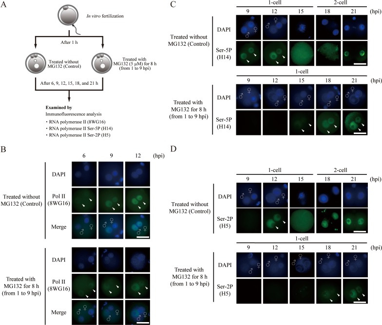
Fig. 2.
Proteasome inhibition affects Pol II localization. (A) Schematic representation of the experimental procedures. (B) Immunofluorescence images of Pol II (green) in untreated and MG132-treated embryos. Representative images of embryos stained for Pol II with anti-8WG16 antibody are shown (arrowheads). (C) Immunofluorescence images of phosphorylation at serine residue 5 (Ser-5P) of Pol II with anti-H14 antibody in untreated and MG132-treated embryos (arrowheads). (D) Immunofluorescence images of phosphorylation at serine residue 2 (Ser-2P) of Pol II with anti-H5 antibody in untreated and MG132-treated embryos (arrowheads). All nuclei were stained by DAPI (blue). Merge images show all images combined with DAPI; ♀, female pronucleus; ♂, male pronucleus; hpi, hours post-insemination; scale bar = 50 µm.

