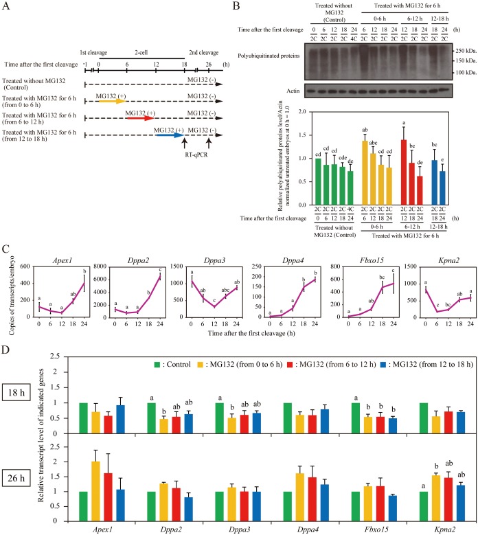
Fig. 4.
Delay of major ZGA by proteasome inhibition. (A) Schematic diagram of experiments in the 2-cell stage. (B) Effect of transient MG132 treatment in 2-cell embryos on the accumulation of polyubiquitinated proteins in embryos. Total proteins isolated from untreated 2-cell (2C), 4-cell (4C), and MG132-treated 2C embryos were immunoblotted with anti-Ub antibody (upper panel). Actin was used as a loading control. Molecular masses (kDa) are shown on the right. Densitometric quantification analysis of the immunoblot bands of polyubiquitinated proteins (lower panel). (C) Expression profile of indicated genes in 2-cell embryos (at 0, 6, 12, and 18 h) and early 4-cell embryos (at 24 h) was confirmed by RT-qPCR. (D) Expression levels in the untreated and MG132-treated embryos at 18 h (upper panel) and 26 h (lower panel). The mRNA levels of the untreated embryos were defined as 1. Untreated (green bars), MG132-treated (from 0 to 6 h) (yellow bars), MG132-treated (from 6 to 12 h) (red bars) and MG132-treated (from 12 to 18 h) (blue bars) embryos; h, time after the first cleavage. Different letters indicate statistical significances (P < 0.05). Bars represent standard error of the mean.

