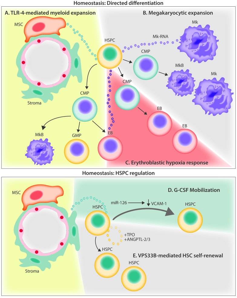
Figure 2.
Current evidence for extracellular vesicle crosstalk in the homeostatic bone marrow microenvironment. (A) MSC-derived EVs signal to HSPCs through the TLR-4 pathway, resulting in myeloid biased expansion. (B) Megakaryocyte-derived MVs are internalized by HSPCs and increase differentiation of new megakaryocytes through RNA-mediated signaling. (C) Hypoxia induces erythroleukemia cells to release EVs containing miR-486 which increases erythroblastic differentiation by targeting Sirt1 in HSPCs. (D) G-CSF infusion stimulates the release of EVs containing miR-126 that act to down-regulate VCAM-1 in HSPCs, resulting in their mobilization out of the BM. (E) HSPCs autoregulate stem potential by packaging and releasing critical secretory proteins through the exosomal pathway via the action of VPS33B. ANGPTL-2/3; angiopoietin-like protein 2 and 3; BM: bone marrow; CMP: common myeloid progenitor; EB: erythroblast; EVs: extracellular vesicles; G-CSF: granulocyte colony-stimulating factor; GMP; granulocyte monocyte progenitor; HSPC: hematopoietic stem and progenitor cell; Mk: megakaryocytes; MkB: megakaryoblast; MSC: mesenchymal stem cell; miR: microRNA; MV: microvesicles; TLR-4: Toll-like receptor 4; TPO: thrombopoietin; VCAM-1: vascular cell adhesion molecule; VPS33B: vacuolar protein sorting-associated protein 33B.

