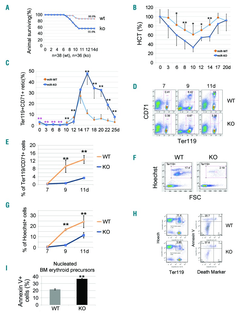
Figure 2.
Apoptotic erythroblasts in miR-144/451−/− bone marrow increase after administration of 5-fluorouracil (5-FU) in adult mice. (A) Survival rates after 5-FU treatment for n=38 (wild-type, WT) and n=36 (miR-144/451−/−, KO) mice. (B) Increased hemolysis of miR-144/451−/− erythrocytes after exposure to 5-FU, as determined by serial hematocrit measurement. N=12 miR-144/451−/− and n=12 WT mice were used. *P<0.05; **P<0.01 (t-test). (C) Flow cytometric analysis of Ter119+CD71+ reticulocytes in circulating blood after 5-FU treatment. N=5 mice of each genotype were used. **P<0.01 (t-test). Note: for the first eight days there were relatively more reticulocytes in miR-144/451−/− blood as compared to WT blood, but significantly fewer during days 8 to 12. The number of reticulocytes in miR-144/451−/− blood dramatically increased around day 14, and much higher levels were sustained than in WT animals. (D) Flow cytometric analysis of Ter119+CD71+ erythroid cells in bone marrow after 5-FU administration for 7–11 days. Note: the appearance of Ter119+CD71+ erythroid cells in bone marrow was delayed relative to that in WT mice, indicating a maturation arrest and/or sustained apoptosis of erythroid cells in miR-144/451−/− mice. (E) Quantitated analysis of Ter119+CD71+ erythroid cells in bone marrow after 5-FU administration for 7–11 days. There were n=5 mice of each genotype at each time point. **P<0.01 (t-test). (F) Flow cytometric analysis of nucleated cell numbers in bone marrow after 5-FU administration. All cells shown in the windows were Ter119+. Gated regions represent nucleated erythroblasts (Hoechst+FSChigh). (G) Quantitative analysis of flow cytometry data from (F). There were n=5 mice of each genotype at each time point. **P<0.01 (t-test). Note: there were far fewer nucleated erythroid cells in miR-144/451−/− bone marrow during days 9 to 11, indicating a maturation arrest and/or sustained apoptosis. (H) Flow cytometric analysis of early apoptosis using Annexin V. Ter119+/Hoechst+ cells are nucleated erythroid cells (left). Near-IR cell death marker−/Annexin V+ cells are early apoptotic cells (right). (I) Quantitative analysis of flow cytometry data from (H). We used n=5 mice of each genotype at day 11 after 5-FU treatment. **P<0.01 (t-test).

