Fig. 2.
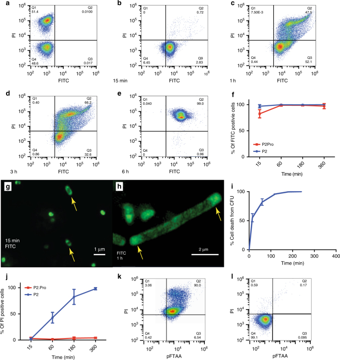
Uptake and inclusion body formation. a–e Fluorescence-activated cell sorting (FACS) analysis of 40,000 E. coli O157: H7 cells, measuring FITC fluorescence (x-axis) and propidium iodide (PI) fluorescence (y-axis) of a untreated and heat-inactivated bacteria mixed at a ratio of 1:1 and b–e bacteria treated for 15 min (b), 1 h (c), 3 h (d), and 6 h (e) with FITC-labeled P2 at MIC concentration. f Average population sizes of FITC-positive cells treated with FITC-P2 or FITC-P2Pro from four independent experiments such as those shown in b–e. g Wide-field structured illumination microscopy (SIM) image of E. coli treated with FITC-P2 for 15 min and h for 1 h at MIC concentration. i Time-dependent cell death following P2 treatment (1 x MIC) as % CFU/mL, in E. coli O157:H7. j Average population sizes of PI-positive cells (propidium iodide) from four independent FACS experiments such as those shown in a–e. k FACS analysis of 40,000 E. coli O157:H7 cells, measuring pFTAA fluorescence (x-axis) and PI fluorescence (y-axis) after 3 h of treatment with P2 at MIC concentration. l Same as h, but after treatment with 100 μg/mL P2Pro

