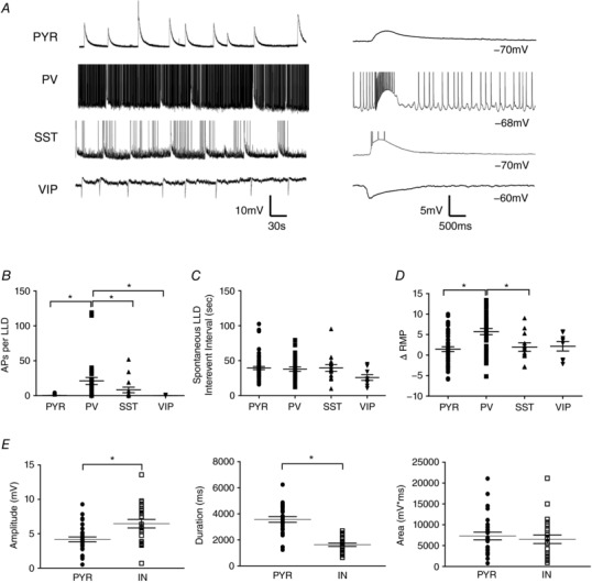
Figure 2. Cell type‐specific properties of spontaneous LLDs.

A, example traces of spontaneous LLDs recorded from a PYR cell and different IN subtypes. Continuously recorded events are shown on the left, with individual events shown on an expanded timescale to the right. LLDs recorded from VIP INs were hyperpolarizing because the RMP of those cells was depolarized relative to the LLD reversal potential. B, cell type comparison of the number of APs superimposed on spontaneous LLDs. C, spontaneous LLDs occurred with the same temporal profile in all cell types. D, application of 4AP + EAA blockers caused a depolarization of all recorded cell types. E, quantitative comparison of spontaneous LLD amplitude (left), duration (middle) and AUC (right) in PYRs and INs. * P < 0.05, Tukey's post hoc test. Each shape represents an individual cell. Error bars are the mean ± SEM.
