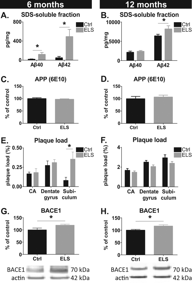
Fig. 1. Hippocampal amyloid pathology in male APPswe/PS1dE9 mice.
a The levels of SDS-soluble Aβ40 and Aβ42 are elevated as a consequence of ELS at 6 months. b At 12 months of age, ELS led to an elevation in Aβ42 levels, but not in Aβ40 levels. c, d No difference was observed between Ctrl and ELS mice for full-length APP levels as measured with 6E10 antibody at 100 kDa at 6 months (c) and 12 months (d). (e) ELS significantly increased amyloid plaque load as measured with 6E10 antibody in the subiculum, but not in the dentate gyrus and cornu ammonis (CA) 1, 2 and 3 areas at 6 months. f ELS did not affect plaque load in the subiculum, dentate gyrus or CA areas at 12 months. g, h BACE1 expression was significantly increased in ELS-APPswe/PS1dE9 mice at 6 (g) and 12 months (h). Six months: Ctrl, N = 5 and ELS, N = 8–10; 12 months: Ctrl, N = 7–9 and ELS, N = 8–9 mice/group

