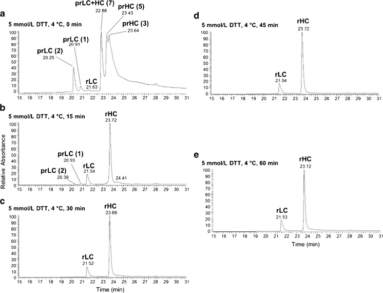
Fig. 2. LC-UV chromatograms of IgG reduced for varying lengths of time.
PS 8670 was reduced at 4 °C in 6.0 mol/L guanidine HCl buffer with 5 mmol/L DTT for (a) 0 min, (b) 15 min, (c) 30 min, (d) 45 min, or (e) 60 min. The level of reduction was determined by LC-UV-MS analysis of 5 μg of mAb. The species comprising each UV peak were identified by deconvolution of their corresponding MS spectra. LC = light chain; HC = heavy chain; pr = partially reduced; r = reduced; the number of intact disulfide bonds is given in parentheses. Representative deconvoluted masses calculated for each species are listed in ESM Table S1

