
Figure 1.
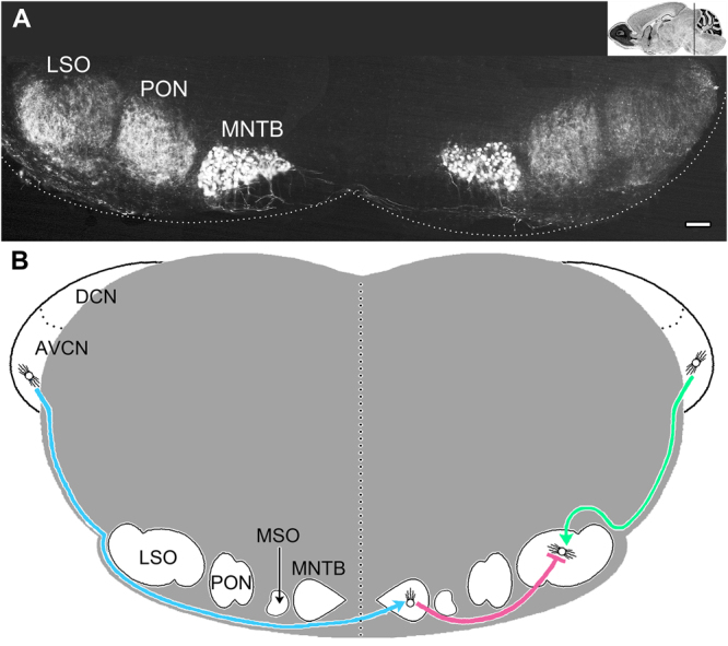
Key nuclei and fiber tracts for binaural transmission in adult mouse auditory brainstem. (A) Calbindin staining labels most of the key bilateral brainstem nuclei in the superior olivary complex (SOC). Principal neurons of the medial nuclei of the trapezoid body (MNTB) are nearest to the midline and form the calyxes of Held. The lateral superior olive (LSO) are the lateral nuclei, and are major signal integrators of binaural signals from the cochleae. The medial superior olive (MSO, not labeled) is poorly developed in mice (approximately 200 neurons) and may only play a minor role in integrating binaural signals. The periolivary nuclei (PON) are likely modulatory but their dispersed cell bodies suggests they generate signals with insufficient synchrony to be detected in far field recordings such as ABRs88. (B) Lower brainstem schematic of three major myelinated fiber tracts and their connections that transmit binaural information to principal cell integrators in the LSO. Only the fiber tracts from the left cochlea to the right LSO (comprising the contralateral pathway), and the right cochlea to the right LSO (the ipsilateral pathway), are shown for clarity. The AVCN – MNTB tract of the trapezoid body (blue) is comprised of large diameter axons and transmits contralateral signals using the excitatory neurotransmitter glutamate; the MNTB – LSO tract (pink) is comprised of intermediate size axons and relays ipsilateral inhibitory signals (glycinergic) in adults; the AVCN – LSO tract (green) is comprised of small axons and transmits excitatory ipsilateral signals (glutamatergic). Each of these nuclei are signal generators for ABR waves. Thus, the cochlear nucleus is associated with wave II, the MNTB with wave III and the SOC with wave IV. Wave V arises from the transmission of signals to the inferior colliculus. Scale bar, 200 μm. Insert in A: Image Credit, Allen Institute.