Figure 3.
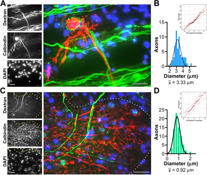
Morphometric analyses of auditory brainstem fiber tracts reveal contralateral axons are large diameter and ipsilateral axons are small. (A) Within the MNTB, a representative contralateral dextran labeled fiber (green) is seen forming the characteristic calyx of Held synapse around a Calbindin + MNTB principal cell (red). Nuclei are stained using DAPI (blue). (B) Distribution of contralateral diameters from dextran-dye labeled axons. The histogram is comprised of 59 axons from 3 mice, 0.2 μm bins, and fit with a normal Gaussian distribution. Mean contralateral fiber diameter is considered large: 3.3 μm. The Q-Q plot (inset) shows most residuals are distributed along the Gaussian diagonal within the 95% confidence envelope (parametric bootstrapping, red dashed lines) as expected for a unimodal distribution. (C) Representative ipsilateral fibers (green) are seen entering the LSO outlined by Calbindin staining (red, white dots). Nuclei are stained using DAPI (blue). (D) Distribution of ipsilateral diameters from dextran-dye labeled axons viewed entering the LSO. Histogram is comprised of 73 axons from 5 mice, 0.2 μm bins, and fit with a normal Gaussian distribution. Mean ipsilateral axon diameter is considered small: 0.91 μm and the Q-Q plot indicates a unimodal distribution. Scale bars, 20 μm.

