Figure 5.
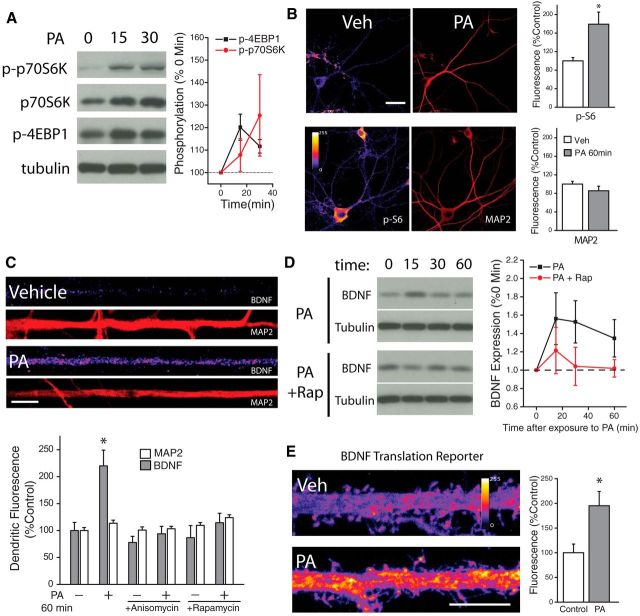
Fast activation of neuronal mTORC1 via exogenous phosphatidic acid. A, Representative Western blots depicting phosphorylated p70S6K (Thr389) and 4EBP1 (Thr37/46) following treatment with PA (100 μm, n = 3 exps) or vehicle for the indicated times; the bands shown are from different blots. Mean (SEM) expression of p-p70S6K and p-4EBP1in neurons subject to treatment with PA. Exogenous PA rapidly enhanced mTORC1 activity, as evidence by significant, time-dependent increases in phosphorylation of its immediate downstream targets p70S6K and 4EBP. 15 min post-PA, n = 3; 30 min post-PA, n = 3; p-p70S6K 15 min post-PA, n = 3; p-p70S6K 30 min post-PA, n = 3. B, Full-frame examples of MAP2 and phosphorylated ribosomal protein S6 (Thr 235/236) staining in neurons treated with vehicle (n = 13) or phosphatidic acid (PA; 100 μm) for 60 min (n = 19). PS6 fluorescence intensity indicated by color look-up table; scale bar equals 40 μm. Mean (+SEM) normalized MAP2 and PS6 expression in neurons treated as indicated. Application of PA activates mTORC1 signaling as indicated by enhanced PS6 staining. *p < 0.05 (t test), relative to vehicle-treated controls. C, BDNF and MAP2 expression in linearized dendritic segments (top), and mean (+ SEM) expression of MAP2 and BDNF in dendritic compartments (bottom) normalized to average control values, in neurons treated with PA (100 μm, 60 min; n = 25) or vehicle (n = 25). BDNF expression in dendrites was significantly (*p < 0.05, relative to vehicle controls) enhanced by treatment with PA compared with vehicle-treated controls. This effect was blockaded by pretreatment with anisomycin (40 μm, 30 min before PA; n = 25) or rapamycin (200 nm, 30 min before PA; n = 25). MAP2 expression did not differ between groups. Scale bar represents 10 μm in A. D, Representative Western blots depicting changes in expression of BDNF or tubulin after treatment with PA (100 μm) ± Rapamycin (300 nm) for the indicated times; the band in the BDNF blot is mature BDNF. Mean (SEM) expression of BDNF in cultured neurons subject to treatment with PA (n = 3 experiments). Exogenous PA rapidly enhanced BDNF expression in an mTORC1-dependent manner, as the increased expression was blocked by rapamycin. E, Representative images of hippocampal neurons expressing myr-d1GFP-nls-A*B treated with either vehicle or PA (100 μm, 45 min). Scale bar, 10 μm. Mean (+SEM) GFP fluorescence in dendrites of hippocampal neurons treated with PA (n = 16) or vehicle (n = 21). Sampled dendritic regions were located 100–200 μm away from the cell body. *p < 0.05 (t test), relative to vehicle-treated controls.

