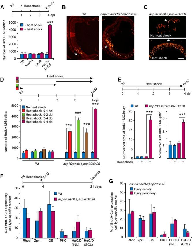
Figure 2.

Forced expression of Ascl1a and Lin28a in the injured retina stimulates MG proliferation and retina regeneration. A, Timeline for experiment and graph showing quantification of BrdU immunofluorescence in INL of injured retinas (needle poke) with forced expression of Ascl1a, Lin28a, or Ascl1a and Lin28a; n = 3 different experiments. Error bars are SD. ***p < 0.001. B, C, Representative images of retinal sections showing BrdU immunofluorescence at the site of injury (needle poke) in Wt and hsp70:ascl1a;hsp70:lin28a transgenic fish with heat shock. Scale bars: B, 150 μm; C, 50 μm. D, Timeline depicting retinal needle poke injury (Inj) followed by a 0–1 hpi, 0–2 dpi, 2–4 dpi, and 0–4 dpi heat shock-treatment and BrdU labeling at 4 dpi. Graph shows quantification of BrdU immunofluorescence in the retina's INL of Wt and hsp70:ascl1a;hsp70:lin28a transgenic fish; n = 3 different experiments. Error bars are SD. ***p < 0.001. E, Timeline of experiment, and graph quantifying the retinal area occupied by BrdU+ MG and their density in Wt and hsp70:ascl1a;hsp70:lin28a transgenic fish ± heat shock. For this analysis a single needle poke injury was made in each retina. Four days later we quantified the area occupied by proliferating MG by counting the number of 12 μm sections spanning the injury site that harbor nine or more BrdU+ MG. Their density per square micrometer was calculated from the three central section's spanning the injury site. Data are normalized to Wt, no heat shock. Error bars are SD. ***p < 0.001; n = 3 different experiments. F, BrdU lineage trace experiment shows forced Ascl1a and Lin28a expression in the injured retina does not bias progenitors toward specific fates. Shown is timeline of experiment and graph quantifying the percentage of BrdU+ cells expressing a particular retinal cell-type marker. Markers are as follows: rods (rho), cones (zpr1), MG (GS), Bipolar (protein kinase C-β1, PKC), amacrine (HuC/D in INL), and ganglion cells (HuC/D in GCL); n = 3 different experiments. Error bars are SD. G, BrdU lineage trace experiment performed as in F shows MG-derived progenitors in the central and peripheral regions of the injury site do not exhibit biases in regenerated cell types when forced to express Ascl1a and Lin28a; n = 3 different experiments. Error bars are SD.
