Figure 6.

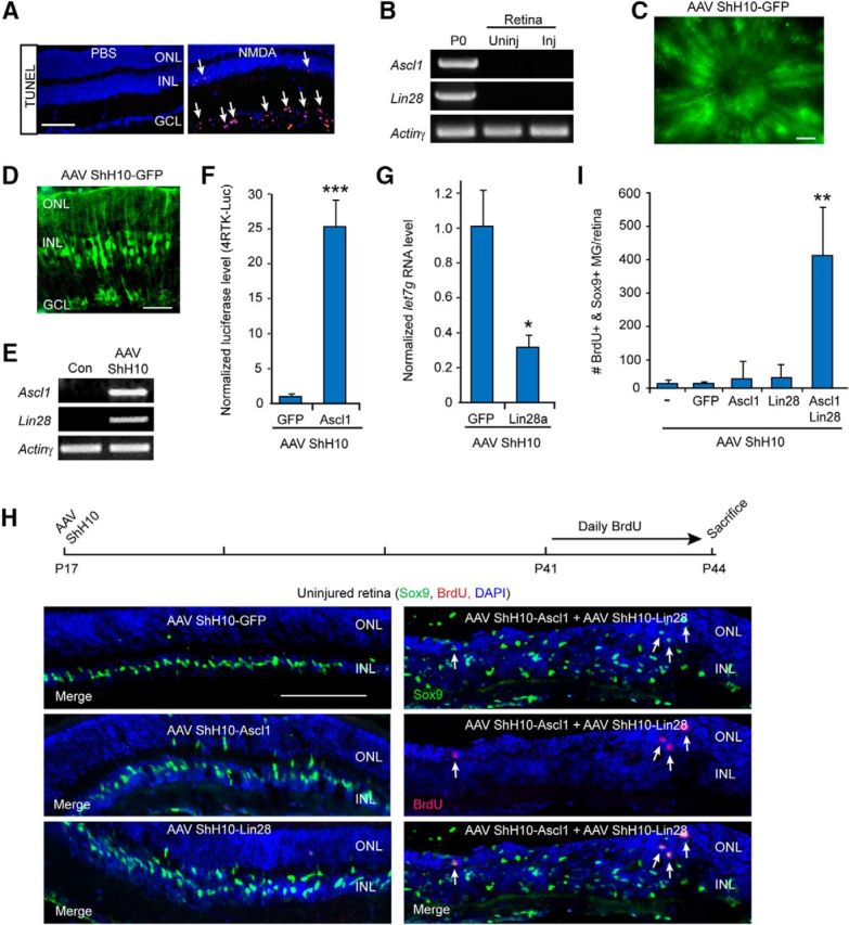
Ascl1 and Lin28a expression synergize with each other to stimulate MG proliferation in the uninjured mouse retina. A, TUNEL stain (red) shows NMDA stimulates cell death in the GCL. Scale bar, 100 μm. B, PCR shows Ascl1 and Lin28a are detectable in the P0 retina and brain, respectively, but not in the uninjured or NMDA damaged (Inj) adult retina. C, GFP fluorescence in flat mount retina shows AAV ShH10-GFP expression throughout the uninjured retina. Scale bar, 100 μm. D, GFP immunofluorescence on retinal sections shows AAV ShH10-GFP expression in MG of the uninjured retina. Scale bar, 50 μm. E, PCR shows Ascl1 and Lin28a expression in retinas transduced with AAV ShH10-Ascl1 and AAV ShH10-Lin28a. F, Luciferase assays show HEK293 cells transfected with 4RTK-Luc and transduced with AAV ShH10-Ascl1 result in increased expression of the 4RTK-Luc reporter indicating expression of a functional Ascl1 protein; n = 3 different experiments. Error bars are SD. ***p < 0.001. G, Transduction of HEK293 cells with AAV ShH10-Lin28a result in reduced let7g expression consistent with expression of a functional Lin28a protein; n = 3 different experiments. Error bars are SD. *p < 0.05. H, Timeline of experiment and representative images of AAV ShH10-Ascl1 and AAV ShH10-Lin28a infected retinas showing Ascl1 and Lin28a synergize to stimulate some MG proliferation (Sox9+/BrdU). Sox9+ MG are labeled green and proliferating BrdU+ cells are labeled red. DAPI labels cell nuclei blue. Scale bar, 100 μm. I, Quantification of data shown in H shows Ascl1 and Lin28a synergize to stimulate MG proliferation in the uninjured retina; n = 4 different experiments. Error bars are SD. **p < 0.01.
