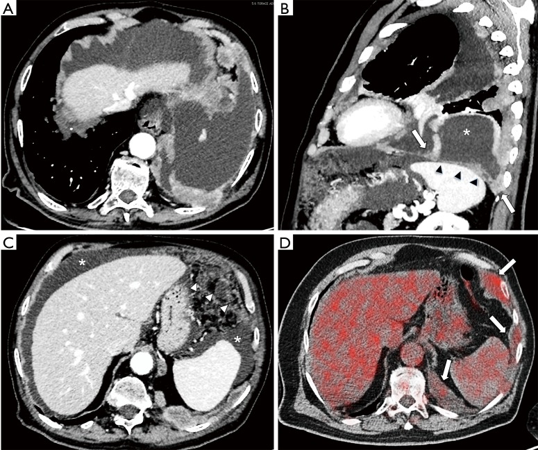
Figure 3.
Trans-diaphragmatic extension in a 62-year-old man with MPM and peritoneal carcinomatosis. (A) Axial contrast enhanced well-collimated multidetector CT (MDCT); (B) sagittal multiplanar MDCT reconstruction images at level of left hemithorax showing nodular pleural thickening in the left hemidiaphragm (arrows) and a left pleural effusion (*). There is complete encasement of left hemidiaphragm with loss of fat plane between diaphragm and spleen (arrowheads) suggestive of transdiaphragmatic extension; (C) MDCT image shows a thick omental thickening in the left anterior abdomen (arrowheads) and ascites (*) due to intraperitoneal neoplastic seeding; (D) axial fused PET/CT image at the level of superior abdomen shows FDG-avid nodular thickening in the left sub diaphragmatic region (arrows).

