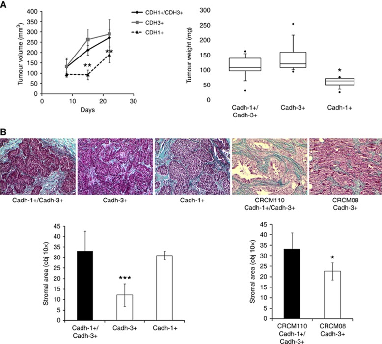
Figure 6.
Effects of cadherin-1 and cadherin-3 silencing on tumour growth and ECM deposition. (A) Different BxPC3 cells lines were injected into the flank of nude mice. For 21 days, mice were monitored weekly for tumour growth (n=6 for each condition). Box plot represents tumour weight 3 weeks after cell inoculation. (B) Tumours were fixed, embedded in paraffin, cut into 4 μm sections, and submitted to Masson’s trichrome staining to distinguish collagen (in blue) from other tissue structures. The stromal area of each tumour was measured by microscopy (× 10 objective) and analysed by ImageJ. *P<0.05, **P<0.01, ***P<0.001.

