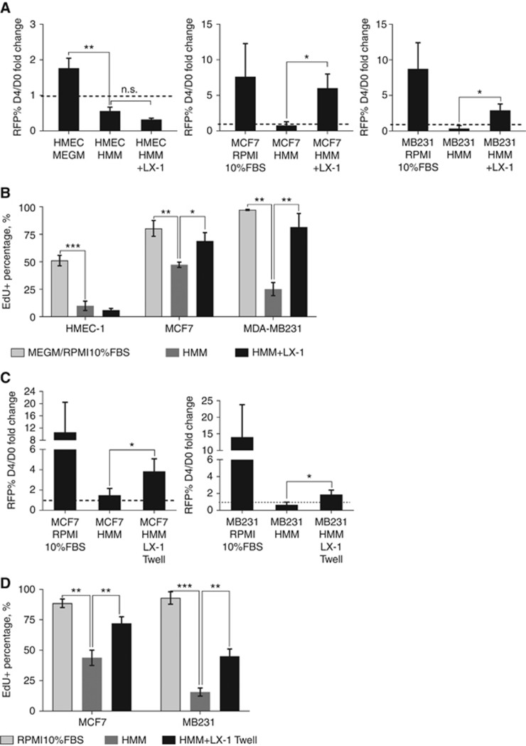
Figure 1.
LX-1 promotes breast cancer cell growth and proliferation in vitro. Serum-free HMM and complete media (RPMI 10%FBS or MEGM) serve as the negative and positive controls, respectively. Average RFP% area fold-change (A) and average EdU incorporation percentage (B) with standard deviation (SD) of normal breast HMEC-1, MCF7 and MDA-MB231 cells in LX-1 co-culture (n=3). Average RFP% area fold change (C) and average EdU incorporation percentage (D) with SD of MCF7 and MDA-MB231 in LX-1 transwell separate culture. n=4 for MCF7 and n=3 for MDA-MB231. *P<0.05, **P<0.01, ***P<0.001.

