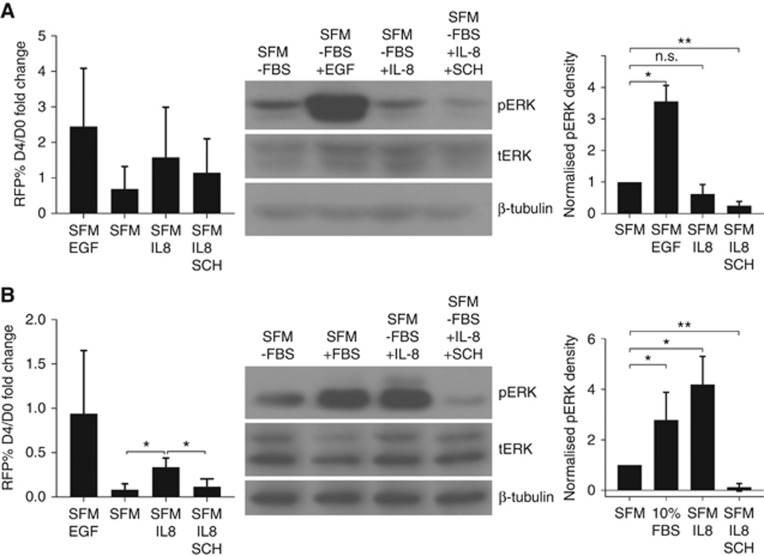
Figure 6.
IL-8 induces ERK phosphorylation. Serum-free RPMI medium (SFM) and SFM+EGF/RPMI+10% FBS serve as the negative and positive controls, respectively. Average RFP% growth fold-change (left panel), immunoblot for phospho-ERK (pERK), total ERK (tERK) and tubulin (middle panel) and pERK density quantification (right panel) of MCF7 (A) and MDA-MB231 (B) cells following 4 days of IL-8 treatments with SD (n=3, n=4 for MCF7 immunoblot). Treatments: 1 μg ml−1 IL-8 for MCF7, 0.25 μg ml−1 IL-8 for MDA-MB231, 20 nM EGF and 0.5 μM SCH772984. *P<0.05, **P<0.01.

