Figure 1.
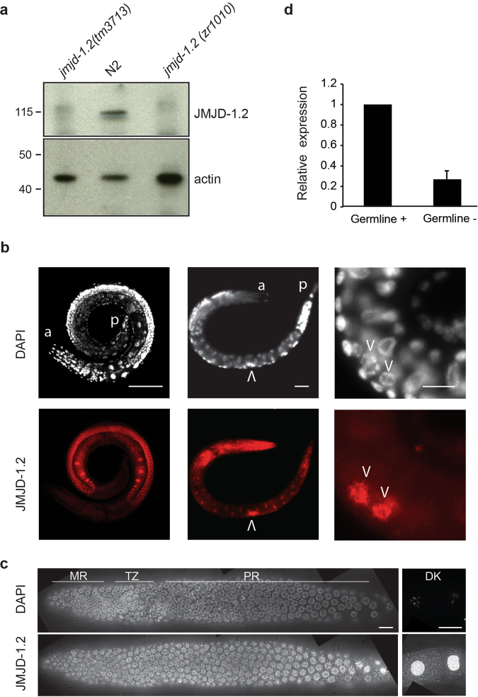
JMJD-1.2 is expressed in the germline. (a) Representative western blot analysis of lysates extracted from the indicated genotypes using JMJD-1.2 antibody. Actin is used as loading control. (b) Representative images of wild-type (N2) animals (adult, left panel; L1 stage, middle and right panels) stained with JMJD-1.2 specific antibody (lower panels) and DAPI staining (upper panels). a, anterior part of the animals, p, posterior part of the animal. Arrowheads indicate the precursor germ cells at L1 stage. Scale bars, 100 μm (left panel) and 10 μm (middle and right panels). (c) Germline excised from N2 young adult hermaphrodite, reconstructed using ImageJ. The mitotic region is on the left and oocytes are in separate panels on the far right. The top panel shows DAPI staining and the bottom panel anti-JMJD-1.2 staining. 100× magnification; scale bar, 10 μm. MR, mitotic region; TZ, transition zone; PR, pachytene region, DK, oocytes in diakinesis. (d) Relative expression of jmjd-1.2 measured by quantitative PCR using glp-4(bn2), grown at 20 °C (gemline+) and at the restrictive temperature of 25 °C (germline −), in which the gonads are absent. cdc-42 is used as internal control. Bar indicates SD from three independent experiments.

