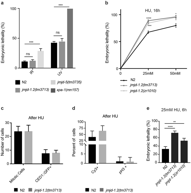
Figure 4.
jmjd-1.2 mutants are hypersensitive to HU. (a) Quantification of the percentage of embryonic lethality in the indicated genotypes in response to radiation (ionizing radiation, IR, 80 Gy; ultraviolet, UV, 300 j/m2). jmjd-5(tm3735) and xpa-1(mn157) mutant alleles are used as positive controls in IR and UV tests, respectively. (b) Quantification of the percentage of embryonic lethality in the indicated genotypes in response to different doses of hydroxyurea (HU) for 16 h. In a and b, the graphics are the average of at least 3 independent experiments and data are presented as mean +/− SEM. ns, no significant differences, *p ≤ 0.05, ***p ≤ 0.001, ****p ≤ 0.0001, comparing the mutant alleles with N2 with paired t-test. (c) Average number of mitotic cells and CED-1::GFP positive cells in N2 and jmjd-1.2(tm3713), grown in 25 mM HU for 16 hours. Data are from at least 15 gonads. (d) Percentage of Cy3-dUTP and pH3 positive mitotic nuclei, in N2 and jmjd-1.2(tm3713), grown in 25 mM HU for 16 hours. Data are from at least 6 gonads and 15 gonads, respectively. In c and d, bars indicate SD. No significant differences were observed with two-tailed paired t-test (p > 0.1). (e) Quantification of the percentage of embryonic lethality in the indicated genotypes in response to exposure to 25 mM HU for 6 hours. The graphic is the average of at least 3 independent experiments and data are presented as mean +/− SEM. **p ≤ 0.01, ****p ≤ 0.0001, with paired t-test.

