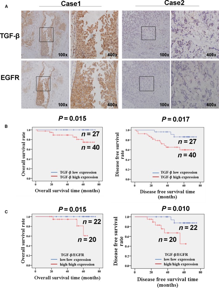
Figure 1.

Elevated expression of TGF‐β is positively correlated with EGFR in breast cancer tissues. (A) The increased staining intensity of TGF‐β is positively correlated with EGFR elevation in invasive ductal breast cancer (IDC). The expression of TGF‐β and EGFR in breast cancer tissue was detected by IHC staining method. Case 1 was synchronously positive expression of TGF‐β and EGFR. Case 2 showed negative expression of both TGF‐β and EGFR. (B) The Kaplan–Meier method was used for survival analysis. The OS and DFS rates in patients with elevated expression of TGF‐β were significantly worse than those in patients with low expression of TGF‐β (OS, P = 0.015; DFS, P = 0.017). The prognosis of the patients with synchronously highly expressed TGF‐β and EGFR was much worse than that of the patients with synchronously poorly expressed TGF‐β and EGFR (OS P = 0.015; DFS P = 0.010).
