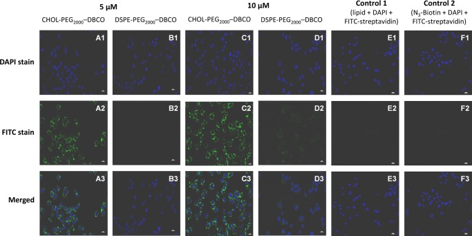
Figure 2.
Confocal microscopy images of RAW 264.7 macrophage cells treated with anchor lipids for 20 min at 37 °C, in phosphate-buffered saline (PBS) buffer (pH 7.4) followed by biotinylation via CFCC and streptavidin-FITC labeling: RAW 264.7 macrophage cells treated with CHOL–PEG2000–DBCO (A, C) and DSPE–PEG2000–DBCO (B, D) with varying concentrations (5 and 10 μM) for 20 min at 37 °C in PBS (pH 7.4) followed by biotinylation and streptavidin-FITC labeling; RAW 264.7 macrophage cells treated with anchor lipids but without azide–PEG4–biotin (E) and treated with azide–PEG4–biotin but without anchor lipids (F).

