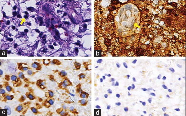
Figure 5.

Histopathological manifestations of patient 4. (a) Hematoxylin and eosin-stained section demonstrating an area of lymphocyte engulfment by a lesional histiocyte consistent with emperipolesis (arrow, original magnification ×400). (b) Immunohistochemical labeling for S-100 protein was diffusely positive within the lesional histiocytes (original magnification ×400). (c) Immunohistological finding: Some histiocytes showed CD68 protein was positive (original magnification ×400). (d) Immunohistochemical labeling for CD1a protein was negative within the lesional histiocytes (original magnification ×400).
