. 2018 Feb 19;8(14):7839–7846. doi: 10.1039/c7ra11943g

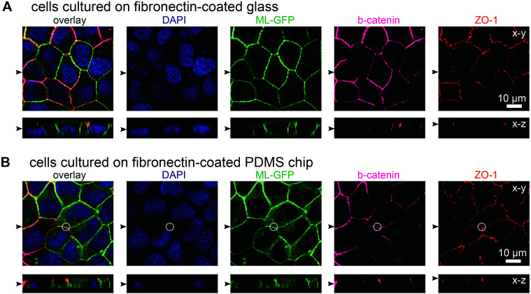
Fig. 2. MDCK cells form polarised monolayers on the biochip. MDCK cells stably expressing ML-GFP (green) were grown for three days on a fibronectin-coated glass cover slip (A) and on a fibronectin-coated PDMS-based biochip (B). After fixation, cells were permeabilised and stained with DAPI highlighting nuclei (blue) and antibodies recognising the basolateral marker beta-catenin (magenta) and the tight junction marker ZO-1 (red). A white circle indicates the perimeter of the pore in (B). Displayed are representative xy sections and xz sections from the acquired confocal image stack. The black arrowheads indicate the positions of the respective image sections.

Fig. 2