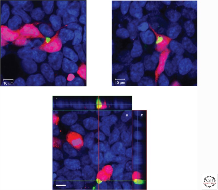
Figure 3.
Intracellular TDP-43 aggregates are transmitted from cell to cell. (Upper) Co-culture of cells expressing DsRed and cells having intracellular TDP-43 aggregates in a 1:1 ratio. After incubation for 3 days, cells were stained with pS409/410 (green) and counter-stained with TO-PRO-3 (blue). Scale bars, 10 μm. (Lower) Cross sections of reconstructed TDP-43 aggregates in these co-cultured cells. (a) Optical section (x-y) at the depth indicated with blue lines in b and c. (b) Cross-sectional y-z image along the green line indicated in a. (c) Cross-sectional x-z image along the red line indicated in a. Scale bar, 10 μm.

