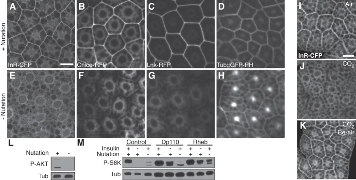
Figure 3.
Localization of insulin signaling components is regulated by mechanical stress. (A–H) Localization of InR-CFP, Chico-RFP, Lnk-RFP, and GFP-PH in fat body cells following 2-h ex vivo incubation of larval carcasses in M3 + insulin medium with (A–D) or without (E–H) nutation. Bar, 20 µm. (I–K) Localization of InR-CFP responds to larval body movement in vivo. CO2 anesthesia of larvae for 15 min led to loss of InR-CFP from the plasma membrane (J) compared with control (I). (K) Normal localization was restored by reaeration of larvae, which allowed recovery of body movement. Bar, 20 µm. (L) The level of phosphorylated AKT, detected by immunoblot of fat body extracts, was elevated by mechanical stress ex vivo. (M) Loss of fb-TOR activity due to lack of insulin in the medium, but not lack of mechanical stress, was partially rescued by fat body-specific overexpression of Dp110 using Cg-Gal4. Overexpression of Rheb in the fat body partially rescued fb-TOR activity in the absence of insulin or mechanical stress.

