Figure 2.

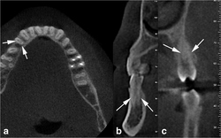
(a, b) Axial and cross-section CBCT image shows showing type V mandibular first premolar. (c) Cross-sectional image showing type II maxillary first premolar.

(a, b) Axial and cross-section CBCT image shows showing type V mandibular first premolar. (c) Cross-sectional image showing type II maxillary first premolar.