
Figure 2.
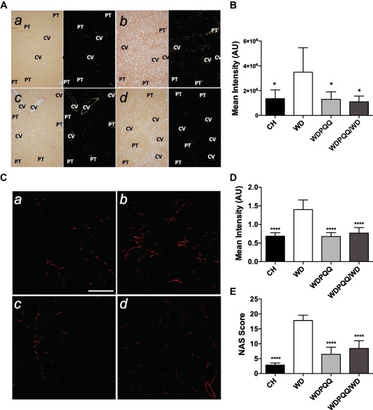
PQQ supplementation diminishes collagen deposition in livers from WD‐fed offspring. (A) Fixed liver sections were stained with picrosirius red and viewed under cross‐polarized light at 100 × to visualize collagen fibrils. a) CH, b) WD, c) WDPQQ, d) WDPQQ/WD. (B) Quantification of positive pixels present in picrosirius red‐stained sections. (C) SHG imaging was performed on fixed liver sections at a magnification of 20×. a) CH, b) WD, c) WDPQQ, d) WDPQQ/WD. (D) Quantification of mean intensity of the SHG signal. (E) Group‐averaged modified NAS, including Brunt criteria, SHG, and CARS indices. Data are mean ± SEM, n = 5‐6/group. Representative images are shown for each group. One‐way analysis of variance with Tukey correction was used to compare between groups. *P < 0.05, ****P < 0.0001 compared to WD. CH, WD, and WDPQQ in A‐E represent both maternal and offspring diets. Abbreviations: AU, arbitrary unit; CV, central veins; NAS, NAFLD activity score; PT, portal triad; SHG, second‐harmonic generation.