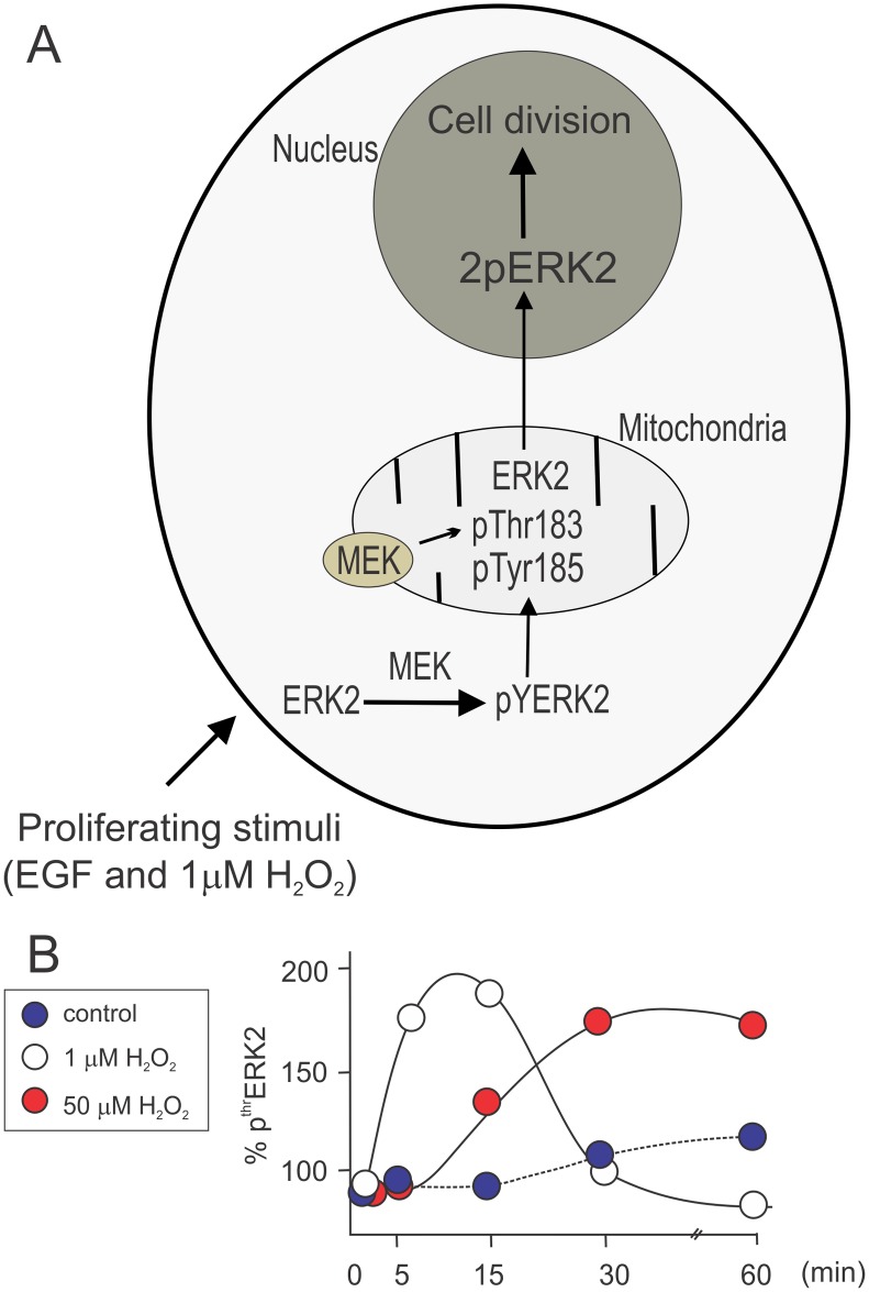
Fig 6. Summary of the proposed mechanistic design for double ERK phosphorylation in cytosol and mitochondria under different redox conditions.
After EGF or 1 μM H2O2 stimulation, which exert a well described and robust proliferative action, ERK2 is rapidly phosphorylated by cytosolic MEK in Tyr185 (pYERK2), allowing a further association with mitochondria and phosphorylation of Thr183, by a mitochondrial MEK pool. This second and compartmentalized phosphorylation drives fully active-ERK2 (2pERK2) translocation to the nucleus to promote gene transcription and cellular division (A). Panel B shows mitochondrial pThr ERK2 kinetics in response to 1 and 50 μM H2O2. At 1 μM H2O2 Thr183 is rapidly phosphorylated in mitochondria with a transient localization and fast translocation to the nucleus (white circles); on the other hand, in the presence of 50 μM H2O2 arresting cell cycle, Thr183 phosphorylated signal is retained in the mitochondrial milieu and ERK translocation to the nucleus is impeded.

