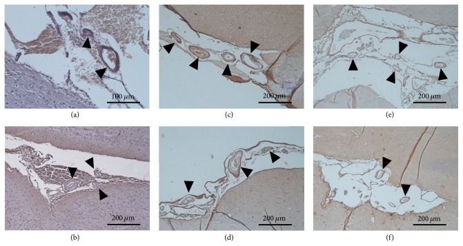
Figure 7.
Cerebral amyloid angiopathy represented by immunohistochemistry (100x magnification). (a-b) Brain section from a young control male and a female animal, respectively. (c-d) Brain section from an aged sedentary male and a female rat, respectively. (e-f) Brain section from an aged running male and a female animal, respectively. The brownish color represents the amyloid-like depositions in the vessel walls.

