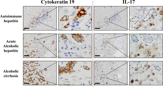
Figure 1.

IL‐17‐expressing cells and CK19+ cells are localized in similar areas in diseased livers. Representative CK19 and IL‐17 (brown color) immunostaining on human serial liver sections from diverse etiologies with an enlargement magnification field. Areas where CK19+ cells accumulate are delimited with dotted lines on serial sections to highlight their proximity with IL‐17+‐infiltrating cells. Scale bar, 100 µm.
