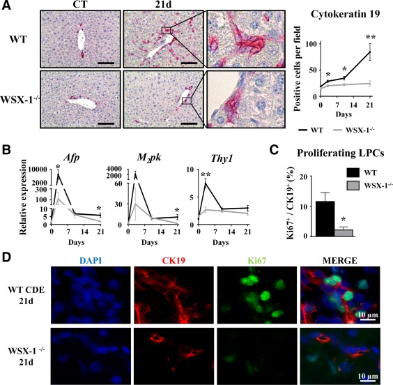
Figure 5.

WSX‐1‐deficiency represses LPC‐driven liver regeneration. Wild‐type and WSX‐1−/− mice were fed a CDE diet, and samples were collected at the indicated time points. (A) CK19+ cells were stained and counted. Scale bar, 100 µm. (B) Hepatic mRNA expressions of LPC‐associated genes were measured by qRT‐PCR. (C,D) CK19 (red) and Ki67 (green) staining and counting after 21 days of the CDE diet. *P < 0.05, **P < 0.01, WT versus WSX‐1−/− mice; each group n = 4‐7 animals. Data represent mean ± SEM. Abbreviations: CT, control; d, day; DAPI, 4′,6‐diamidino‐2‐phenylindole; M2pk, type 2 muscle pyruvate kinase; qRT‐PCR, quantitative reverse‐transcription polymerase chain reaction.
