Figure 6.

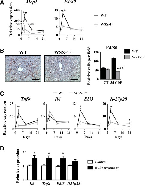
CDE diet‐induced liver inflammation is reduced in WSX‐1−/−. Wild‐type and WSX‐1−/− mice were subjected to the CDE model. (A) Hepatic mRNA expression of macrophage‐related genes was assessed by qRT‐PCR. (B) Immunostaining of F4/80 was performed on WT and WSX‐1−/− mice, and positive cells were counted after 3 days of the CDE diet. Scale bar, 100 µm. (C) Hepatic mRNA expressions of inflammation‐related genes were quantified by qRT‐PCR; each group n = 4‐7 animals. *P < 0.05, **P < 0.01, ***P < 0.005, WT versus WSX‐1−/− mice. (D) RAW264.7 cells were cultured in the presence of 50 ng/mL IL‐27, and mRNA expressions of IL‐6, TNF‐α, and IL‐27 subunits were analyzed by qRT‐PCR. *P < 0.05, control versus IL‐27‐treated cells. Data represent mean ± SEM. Abbreviations: CT, control; d, day; qRT‐PCR, quantitative reverse‐transcription polymerase chain reaction.
