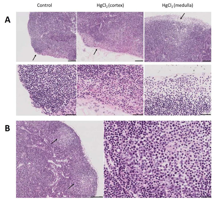
Figure 2.
Development of germinal centers in draining lymph node following HgCl2 exposure. A, Popliteal lymph node 3 hours after footpad injection of HgCl2 showing marked enlargement of the subcapsular sinus and large numbers of polymorphonuclear leukocytes (arrows). Similar areas were selected for the control and HgCl2 (cortex) images. Notice that the subcapsular sinus in the control is barely visible below the single cell layer capsule. B, Popliteal lymph node 7 days after footpad injection of HgCl2 showing germinal centers (GC) (left panel, arrows). An enlargement of the GC area is shown in the right panel. Bars in A are 100 μm in top panel and 50 μm in the bottom panel. In B bar on the left is 100 μm and 25 μm on the right.

