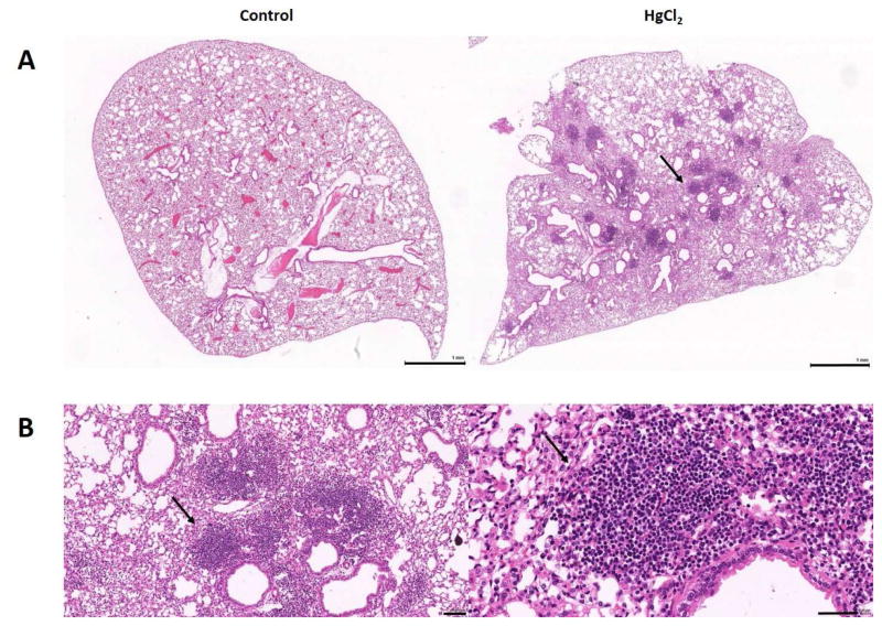
Figure 3.
Mercury-induced ectopic lymphoid structures (ELS) in the lung. A, Lungs from mice given PBS (Control) or HgCl2 showing the presence of lymphoid cell accumulations (arrow) after mercury exposure for 4 weeks. B, Enlarged views of the area indicated by the arrow in A showing the dense accumulation of lymphoid cells. Bars in A are 1 mm and in B 100 μm on the left and 50 μm on the right.

