Figure 2. Mesenteric and hepatic tumors in 18 month old K1 transgenic mice.
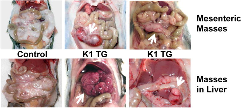
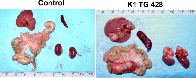
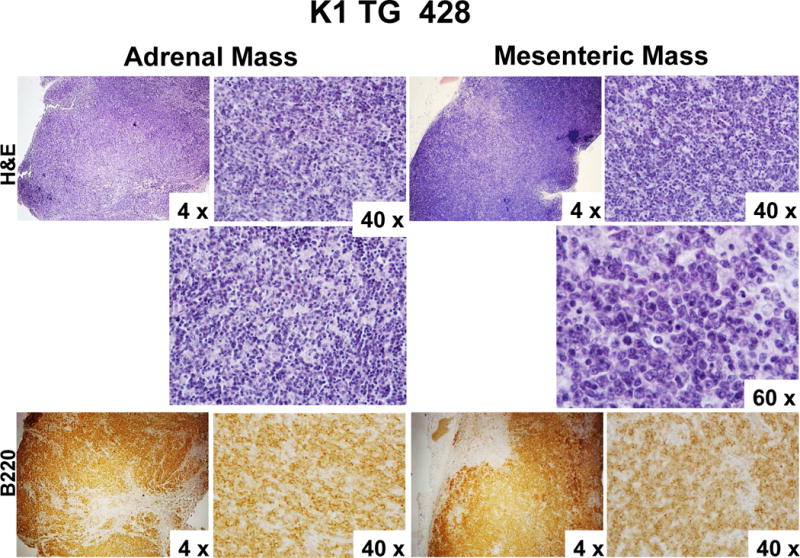
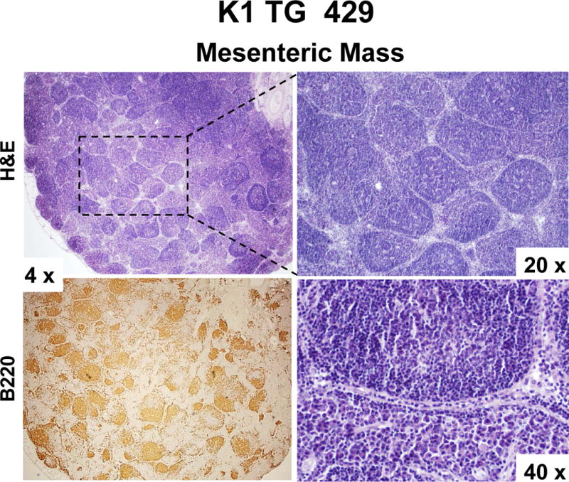
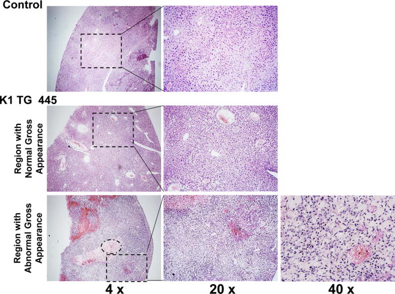
(A) Gross examination of two control (K1-negative littermates) and four K1 transgenic mice with tumors/masses in the mesentery and livers; (B) Harvested livers, intestines, spleens and kidneys from control (K1-negative littermate) and K1 transgenic mouse # 428 with tumors/masses in the mesentery, liver, and adrenal glands; (C) Hematoxylin/eosin (H&E) and B-cell marker B220 staining of adrenal and mesenteric masses isolated from K1 transgenic mouse # 428; (D) H&E and B-cell marker B220 staining of mesenteric mass isolated from K1 transgenic mouse # 429; (D) H&E staining of livers from control (K1-negative littermate) and K1 transgenic mouse # 445 showing regions with an abnormal appearance.
