
Extended Data Figure 11. Different response to CDX2 overexpression in the transitional (p63+ KRT7+) and the squamous (p63+ KRT7−) basal progenitor cells in vitro. Human and mouse MLE present similar gene expression.
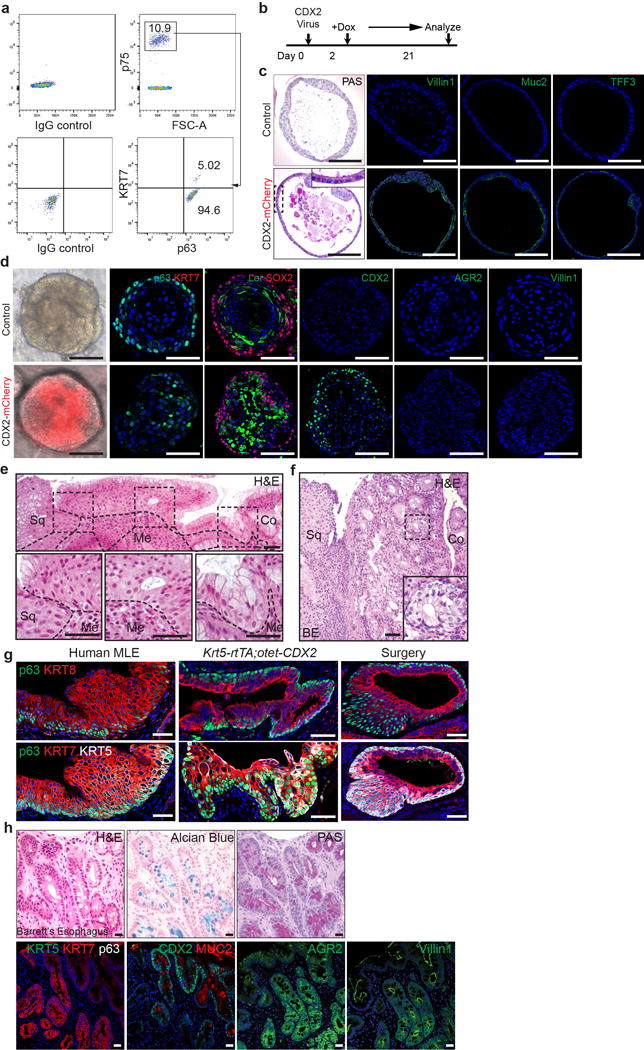
a, Two distinct basal progenitor populations (p63+ KRT7− Vs p63+ KRT7+) are present at the human SCJ as indicated by flow cytometric analysis. n=3 independent experiments. b, Schematic depicts the induction of CDX2 overexpression with Doxycycline treatment of CDX2 virus-infected human SCJ basal progenitor cells. c, CDX2 overexpression promotes intestinal metaplasia of p63+ KRT7+ cells. The metaplastic columnar cells are PAS+ and express Villin1, Muc2 and TFF3. n=6 per group. d, Ectopic CDX2 expression does not promote intestinal metaplasia of the stratified squamous epithelium in organoids formed by p63+KRT7− squamous basal cells. n=4 per group. e, The transitional epithelium with underlying basal cells is dramatically expanded in patients with long-term gastro-esophageal acid reflux. Dotted lines indicate the basement membrane. n=3. f, The transitional epithelium with basal cells is amplified in BE mixed with MLE. n=5. g, Similar phenotypic presentation of human MLE and mouse MLE developed at the SCJ following CDX2 overexpression and oesophageal-duodenal anastomosis surgery. human MLE n=10; Krt5-rtTA; otet-CDX2 mutants n=5; surgical mice n=5. h, Goblet cells in human BE is positive for Alcian blue and PAS staining. BE epithelium loses the expression of KRT5 and p63 while maintaining the expression of KRT7. Note BE gains the expression of CDX2, MUC2, AGR2 and Villin1. n=12. Scale bar, 20 μm.