
Extended Data Figure 2. Expansion of the columnar epithelium at the SCJ in Krt5-CreER;R26Sox2-GFP mutants.
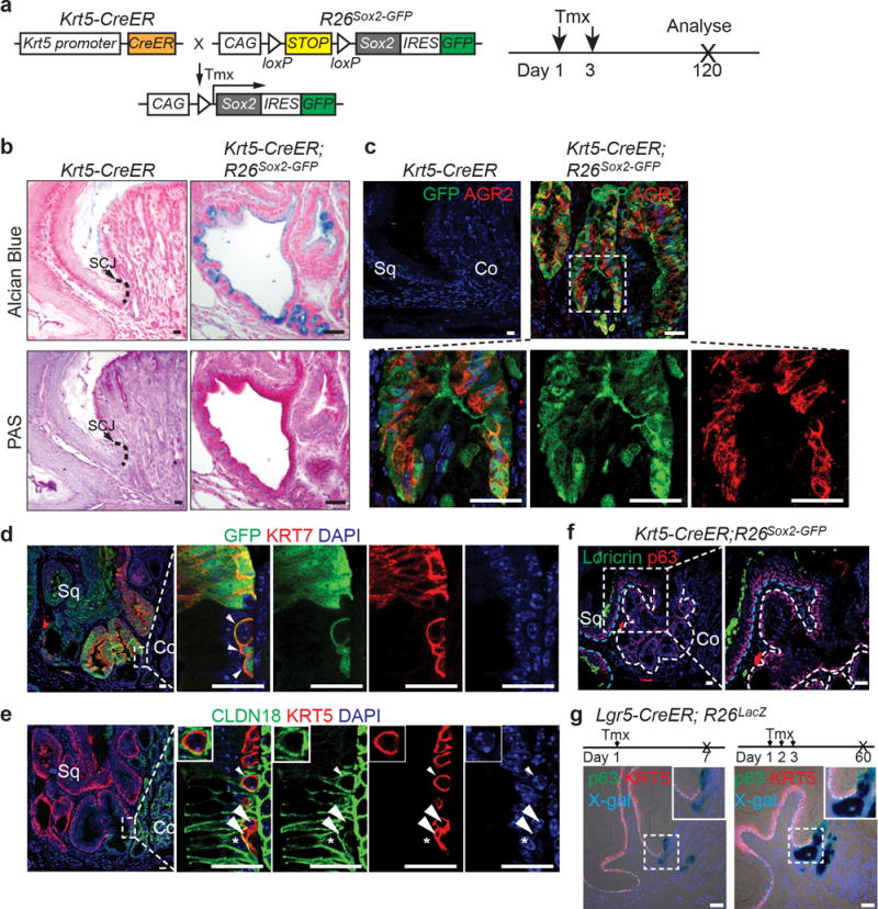
a, Schematic depicts generation of Krt5-CreER;R26Sox2-GFP (SOX2 overexpression) mutants. b, The columnar epithelium secretes mucin as indicated by Alcian blue and periodic acid-Schiff staining. n=7 per group. c, The mucin secreting cells (AGR2+) are derived from KRT5+ basal progenitor cells as verified by the lineage tracing tag GFP. n=7. d, High magnification picture of Figure 1b to show that expanded GFP+KRT7+ basal progenitor cells invade underneath of the cardia mucosa upon SOX2 overexpression. n=7. e, High magnification picture of Figure 1c to show that expanded basal cells (KRT5+) invade and intercalate with the cardia mucosal epithelium (CLDN18+) upon SOX2 overexpression. Note KRT5+ cells (arrow and arrowheads) do not express CLDN18. Conversely, CLDN18+ cell does not express KRT5 (star). n=7. f, The columnar epithelium does not express the squamous cell marker Loricrin. n=7. The white and blue dotted lines indicate the amplified columnar epithelium and the stratified squamous epithelium, respectively. g, Co-staining of X-gal with KRT5 and p63 indicates Lgr5+ cardia progenitor cells do not contribute to KRT5+ or p63+ basal cells in Lgr5-CreER; R26lacZ mice with both short-term tracing and long-term tracing. n=3 per group. Scale bar, 20 μm.