
Extended Data Figure 3. Basal progenitor cells contribute to columnar metaplasia following anastomosis surgery-induced bile acid reflux.
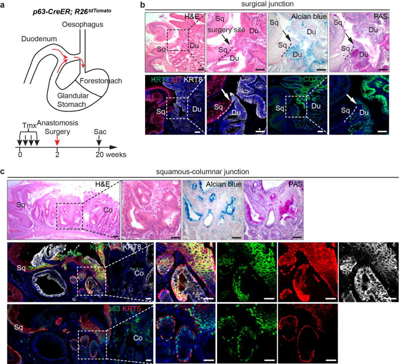
a, Schematic depicts the use of oesophageal-duodenal anastomosis and lineage tracing in p63-CreER;R26tdTomato mice. b, Metaplasia does not occur at the distal oesophagus where the anastomosis surgery site is located (arrow). Note that Alcian blue, PAS and CDX2 label the intestinal but not the squamous oesophageal epithelium. n=5. c, Lineage-labeled transitional epithelial cells (tdT+) expand and resemble the multilayered epithelium, expressing the columnar markers (KRT7, KRT8) and basal cell markers (KRT5 and p63). The expanded epithelium secretes mucin as indicated by Alcian blue and PAS staining. n=5. Scale bar, 20 μm.