
Extended Data Figure 5. Loss of p63 prevents the stratification of KRT7+ columnar epithelium during embryonic development.
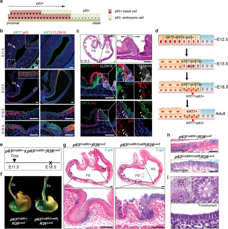
a, Diagram shows the proposed REC model of downward expansion of p63+ KRT7− basal cells and retreatment of p63−KRT7+ embryonic cells. b, The columnar epithelium lining the mouse forestomach and the SCJ expresses p63, KRT7 and KRT8 from E11.5 to E16.5. Note Claudin18 expression is limited to the hindstomach epithelium. n=5. c, Expression of KRT7 is restricted to the SCJ transitional epithelium at E18.5, and basal cells (arrowheads) express p63, KRT5, KRT7 and low levels of KRT8 but not CLDN18. n=5. d, Schematic depicts the gradual restriction of KRT7 expression to the SCJ transitional epithelium during development. Initially the simple columnar epithelium lining the forestomach and SCJ expresses both p63 and KRT7. Upon stratification the forestomach epithelium loses KRT7 expression, while basal cells at the SCJ maintain expression of both p63 and KRT7. e, Lineage tracing of epithelial progenitor cells (p63promoter active) in p63CreER/CreER; R26lacZ (p63 null) mutants. f, Whole-mount x-gal staining of the oesophagus and stomach isolated from p63CreER/CreER; R26LacZ mutants and p63CreER/+; R26LacZ controls. n=3. g, h, The simple columnar epithelium lining the forestomach (g) and oesophagus (h) of mutants is derived from basal progenitor cells (p63 promotor active) as indicated by x-gal staining. n=3 per group. i, Normal oesophagus and forestomach is lined by simple columnar epithelium at E11.5. n=3. Abbreviation: Es, oesophagus; FS, forestomach; HS, hindstomach; Tr, trachea. Scale bar, 20 μm.