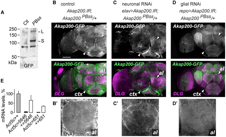
Figure 2. Akap200 Is Neuronal and Glial.

(A) Western blot of Akap200-GFP protein trap and control extracts probed with GFP antibodies.
(B) Akap200-GFP is expressed broadly in the adult brain, labeled with GFP (green) and DLG (magenta) antibodies. ctx: cortex region containing glia and neuron cell bodies; al: antennal lobe. Genotype: w,UAS-Akap200.IR/+;Akap200PBss/+. Scale bar: 50 μm.
(B′) Akap200-GFP in the antennal lobe.
(C) Akap200 RNAi in all neurons reduced synaptic neuropil Akap200-GFP.
(C′) Akap200-positive ensheathing glia. Genotype: w,elav(c155)-Gal4,UAS-Akap200.IR/+;Akap200PBss/+.
(D) Akap200 RNAi in all glia reduced Akap200-GFP expression.
(D′) Loss of glial staining in the antennal lobe. Genotype: w,UAS-Akap200.IR/+;Akap200PBss/+;repo-Gal4/+.
(E) Akap200 qPCR for all transcripts in fly heads when Akap200 RNAi is expressed ubiquitously with Act5c-Gal4, normalized to Act5c > +. All bar graphs are mean with SEM.
