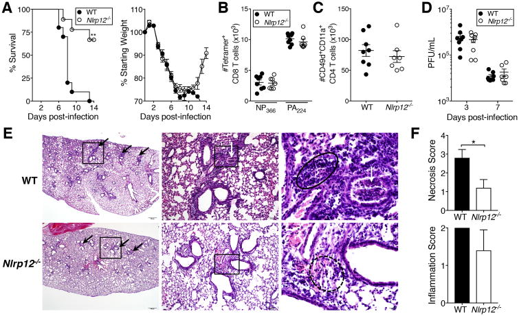
Figure 1. Improved survival of Nlrp12-/- mice following lethal IAV infection.
(A) Mice were infected with a 4LD50 inoculum of IAV and monitored for survival and weight loss through day 14 post-infection. (B, C) Seven days post-infection, IAV-specific CD8+ (B) and CD4+ (C) T cells in the lungs were enumerated using indicated markers. (D) At days three and seven post-infection, virus was quantified in lung homogenate supernatants by plaque assay. (E) H&E-stained sections of lungs harvested five days post-infection. Scale bars and original magnification: left, 500μm (2×); middle, 100μm (10×); right, 20μm (50×). Boxes indicate the area magnified in the panel immediately to the right. Black arrows indicate peribronchiolar inflammation, white arrows indicate cellular debris within the airway, solid circle outlines lymphocytic infiltrates, dotted circle outlines scattered mixed inflammatory cells within the pulmonary interstitium. (F) Scoring of necrosis and inflammation in H&E stained lung sections. (A) Results are representative of two independent experiments, n=10 WT, 9 Nlrp12-/-; (B-D) results are representative of two independent experiments, n= 6-8 per group; (E, F) results are representative of one experiment, n=5 per group, graphed as mean ± SD. *p<0.05, one sample t-test; **p<0.01, Gehan-Breslow-Wilcoxon test.

