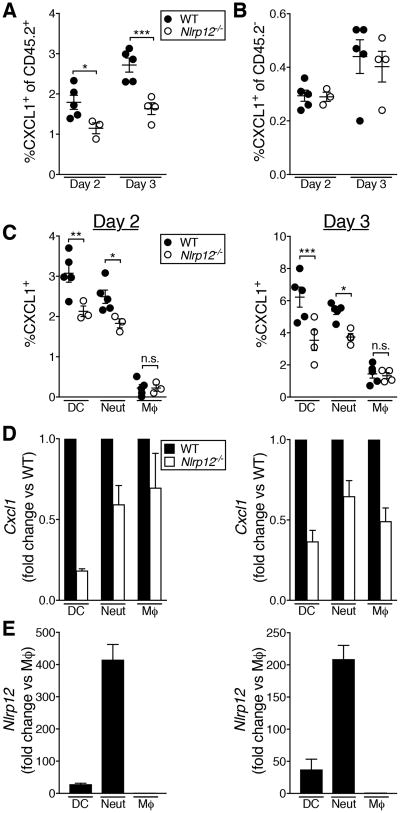
Figure 6. Decreased CXCL1 production by immune cells in the lungs of Nlrp12-/- mice during IAV infection.
Mice were infected with a 4LD50 inoculum of IAV. (A-C) At the indicated day post-infection, CXCL1+ cell populations were enumerated in the lungs by flow cytometry. (D, E) At the indicated day post-infection, cell populations were purified by FACS and gene expression was determined by qRT-PCR. (A-C) Results are representative of two independent experiments, n=3-5 per group, error bars represent SEM. (D, E) Data were pooled from two independent experiments, graphed as mean ± SEM * p<0.05, ** p<0.01, *** p<0.001, student's t-test.

