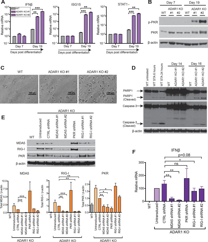
Figure 7. Differentiation of ADAR1 KO hESCs to NPCs leads to spontaneous IFNβ production, PKR activation, and apoptotic cell death.
(A–D) WT and two independent ADAR1 KO hESC clones were differentiated into monolayer NPCs (Figure S7A). qRT-PCR of IFNβ and two ISGs (STAT1, ISG15) (n = 3 experimental replicates) (A). Western blot of p-PKR in NPCs (B). Bright field images of NPC monolayer at 20 days post differentiation (C). Western blot of full length and cleaved forms of PARP1 and Caspase-3, markers of apoptosis (D). As a positive control for apoptosis, WT NPCs were treated with 1uM of Staurosporine (STA).
(E-F) RIG-I, MDA5, or PKR were knocked down in ADAR1 KO NPCs with lentiviral shRNAs (Figure S7F). Representative western blot (uppler panel) and band intensities normalized to β- actin (lower panels, n = 2~5 experimental replicates) (E). qRT-PCR of IFNβ mRNA (n = 4~5 experimental replicates) (F).
(A, E, F) Data shown as mean ± SEM. Student’s t-test, *P < 0.05, **P < 0.01, ***P < 0.001, ns = not significant.
See also Figure S7.

