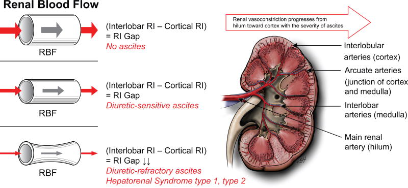
Figure 2.
Renal vasoconstriction in cirrhosis progresses from main renal artery (hilum), toward interlobar arteries (renal medulla) and finally affects arcuate (junction of renal medulla and cortex) and interlobular arteries (renal cortex).40 There is an inverse relationship between renal blood flow (RBF) and renal resistive index (RI).39, 40 When RBF decreases, renal RI increases. 39, 40 While patients without ascites and with diuretic-sensitive ascites preserve cortical renal blood, those with diuretic-refractory ascites have a substantial reduction in cortical renal blood flow. 39, 40 Therefore, while there is a renal RI gap between interlobar and cortical arteries in patients with cirrhosis without ascites and with diuretic-sensitive ascites, this RI gap disappears due to an increase in both interlobar and cortical RIs in patients with cirrhosis and diuretic-refractory ascites.40 Cortical ischemia is considered to be the landmark feature of cirrhosis and diuretic-refractory ascites and HRS.1, 38-41 (Used with permission of Baylor College of Medicine).

