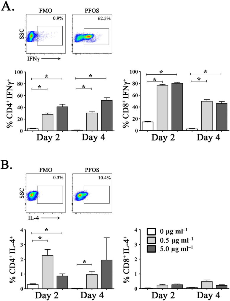
Figure 3.
In vitro exposure to PFOS induces potent IFNγ production from dolphin CD4+ and CD8+ T cells. Production of (A) IFNγ and (B) IL-4 in CD4+ (left) and CD8+ (right) T cells determined by intracellular cytokine staining following in vitro culture with 0 µgml−1 (open bars), 5 µgml−1 (gray bar), or 50 µgml−1 (dark bars) exogenous PFOS. Representative images of dolphin peripheral blood T cell production of IFNγ (CD8+) and IL-4 (CD4+) following in vitro stimulation with exogenous PFOS. Representative FMO-negative gating controls are illustrated for IFNγ and IL-4 staining. *P ≤ 0.001. FMO, fluorescence minus 1; IFNγ, interferon-γ; IL-4, interleukin-4; PFOS, perfluorooctane sulfonate; SSC, side scatter. [Colour figure can be viewed at wileyonlinelibrary.com]

