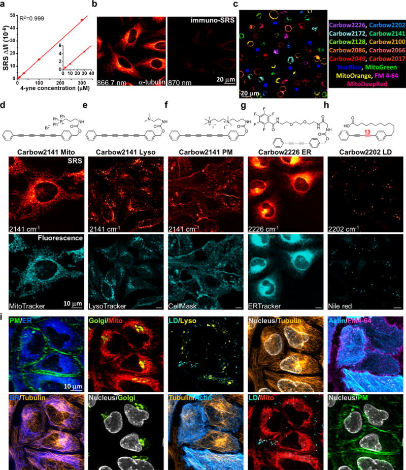
Figure 4. Super-multiplexed optical imaging with polyynes.

(a) Linear concentration dependence of 4-yne with sub-μM SRS detection sensitivity. Error bars: mean ± s.d., n=3 measurements. Solid line shows a linear fitting (R2=0.999, n=5 concentrations). (b) Immuno-staining and imaging of α-tubulin in fixed HeLa cells with 4-yne conjugated antibody. (c) 15-color tandem fluorescence-SRS imaging of live HeLa cells with corresponding Carbow and fluorescent molecules. (d-h) Chemical structures of five organelle-targeted probes based on polyynes for live-cell imaging, including mitochondria Mito (d), lysosome Lyso (e), plasma membrane PM (f), endoplasmic reticulum ER (g) and lipid droplet LD (h). (i) 10-color optical imaging of PM (Carbow2141), ER (Carbow2226), Golgi (BODIPY TR), Mito (Carbow2062), LD (Carbow2202), Lyso (Carbow2086), nucleus (NucBlue), tubulin (SiR650), actin (GFP) and FM 4-64 in living HeLa cells. Overlay of two species are shown in each image.
