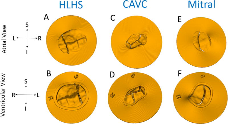
Figure 4.

Models Ready for Direct 3D Printing from atrial and ventricular viewpoints: (A-B) Tricuspid in HLHS; (C-D) CAVC; (E-F) Mitral. Note preservation of coaptation with a small gap artificially created to ensure the ability to separate leaflets after printing. The coaptation gap for the tricuspid and mitral was 0.3mm and 0.2mm for the mitral.
