
Figure 2.
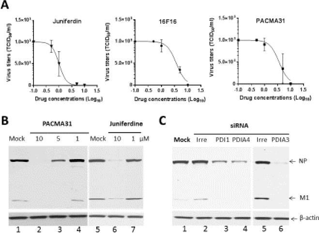
Effects of PDI inhibitors or knockdown of PDI1, PDIA3 or PDIA4 expression by siRNAs on the replication of influenza virus in MDCK cells. A) Dose-response curves of the antiviral activities of Juniferdin, 16F16 or PACMA31. Confluent MDCK cells were infected with influenza A virus, A/WS/33, at an MOI of 0.1 in the presence of Juniferdin, 16F16 or PACMA31 at various concentrations or mock-medium for 1hr. Then, the cells were washed with phosphate buffered saline (PBS), and fresh media containing trypsin (2 μg/ml) and the compound or mock-medium was added to the cells. Virus titers were determined at 2 days post infection using the TCID50 method. B) Expression of influenza virus proteins in the presence of various concentrations of PACMA31 or Juniferdine. Cells were infected with A/WS/33 at an MOI of 5 MOI and incubated in the presence of mock-medium or various concentrations of PACMA31 or juniferdine for 24 hrs. C) Effects of knockdown of PDI1, PDIA3 or PDIA4 expression by siRNAs on the replication of influenza virus in MDCK cells. Cells were mock-transfected or transfected with irreverent RNA or siRNAs targeting each gene. At 24 hr of transfection, cells were infected with A/WS/33 at an MOI of 5. Western blot analysis of cell lysates in B and C was performed with antibodies against whole influenza virus or β-actin. Viral NP and M1 proteins and β-actin are shown.