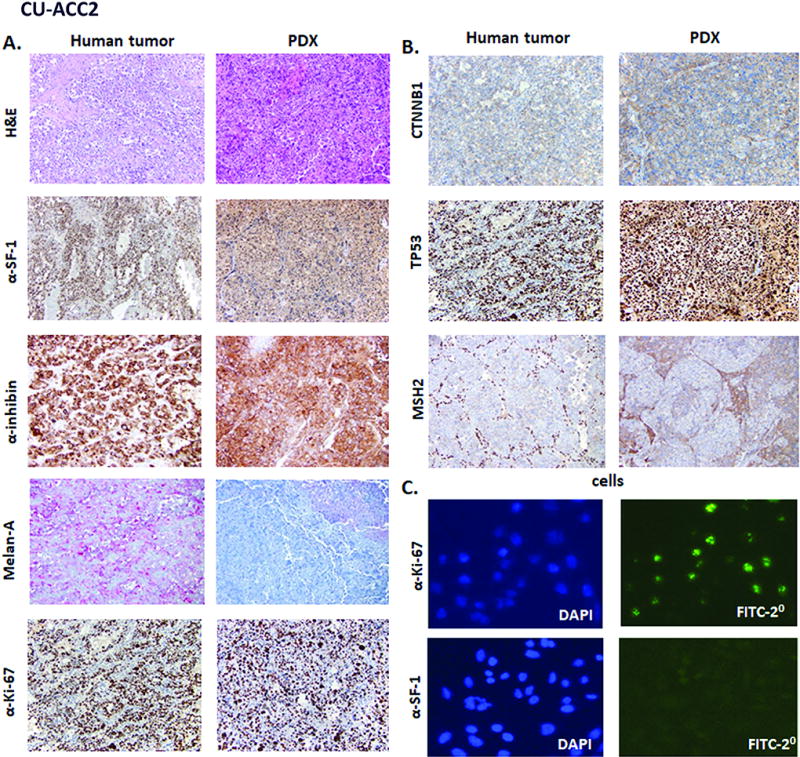
Figure 3.

Immunohistochemistry of human CU-ACC2 tumor and CU-ACC2 PDX. A. and B. The left columns shows CU-ACC2 human tumor sample and the right two column is from CU-ACC2 PDX. A. The immunochemistry stains include H&E, SF1, α-inhibin, Melan-A, Ki-67 and B. β-catenin, p53 and MSH2 C. Immunocytochemistry for Ki67 and SF1 (right columns) for CU-ACC2 cells (DAPI left column).
