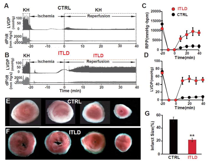
Figure 2. Administration of ITLD during reperfusion improves heart functional recovery and reduces myocardial infarct size of LP mice subjected to ex-vivo I/R injury.
A, B. Representative traces of the left ventricular pressure and heart contractility as a function of time in CTRL and 1% ITLD group, respectively. C. Rate Pressure Product (RPP) and D. left ventricular developed pressure (LVDP) as a function of time in CTRL and ITLD. E, F. Four slices of the same heart after TTC staining in CTRL and ITLD, respectively. The white area represents the infarct zone and red shows the viable area. G. The area of necrosis as the percentage of total left ventricular (LV) area in CTRL and ITLD. *p<0.05 and **p< 0.01 vs. CTRL (n=7–8).

